Difference between revisions of "General Information/Transposase expression and activity"
| Line 41: | Line 41: | ||
Transposase stability can also contribute to control of transposition activity. The Tpase of IS''903'' is sensitive to the ''E. coli'' Lon protease<ref><nowiki><pubmed>2161528</pubmed></nowiki></ref>. This sensitivity limits the activity of the Tpase both temporally and spatially and may provide an explanation for the observation that several Tpases function preferentially in cis. Indeed mutant IS''903'' Tpase derivatives have been isolated which exhibit an increased capacity to function in trans. These are more refractory to Lon degradation than the wildtype protein<ref><nowiki><pubmed>8898394</pubmed></nowiki></ref>. Some evidence that Lon may also be involved in regulating Tn''5'' (IS''50'') transposition has also been presented<ref><nowiki><pubmed>2438419</pubmed></nowiki></ref>. | Transposase stability can also contribute to control of transposition activity. The Tpase of IS''903'' is sensitive to the ''E. coli'' Lon protease<ref><nowiki><pubmed>2161528</pubmed></nowiki></ref>. This sensitivity limits the activity of the Tpase both temporally and spatially and may provide an explanation for the observation that several Tpases function preferentially in cis. Indeed mutant IS''903'' Tpase derivatives have been isolated which exhibit an increased capacity to function in trans. These are more refractory to Lon degradation than the wildtype protein<ref><nowiki><pubmed>8898394</pubmed></nowiki></ref>. Some evidence that Lon may also be involved in regulating Tn''5'' (IS''50'') transposition has also been presented<ref><nowiki><pubmed>2438419</pubmed></nowiki></ref>. | ||
| − | An observation which might also reflect Tpase instability is the temperature sensitive nature of IS''1''-mediated adjacent deletions ''in vivo''<ref><nowiki><pubmed>1101028</pubmed></nowiki></ref>, of Tn''3'' transposition<ref><nowiki><pubmed>378977</pubmed></nowiki></ref> and of IS''911'' intramolecular recombination both in vivo and in vitro<ref><nowiki><pubmed>9302015</pubmed></nowiki></ref>. For IS''911'', incubation of the Tpase at 42°C results in an irreversible loss in activity. Further analyses showed that there was an increase in the proportion of transposase fragments some of which can presumably interact with full length transposase to inhibit its activity. This effect can be somewhat reduced by a series of mutation in the transposase gene whose function in stabilising the transposase is as yet unknown<ref><nowiki><pubmed>17078817</pubmed></nowiki></ref>. | + | An observation which might also reflect Tpase instability is the temperature sensitive nature of IS''1''-mediated adjacent deletions ''in vivo''<ref><nowiki><pubmed>1101028</pubmed></nowiki></ref>, of Tn''3'' transposition<ref><nowiki><pubmed>378977</pubmed></nowiki></ref> and of IS''911'' intramolecular recombination both ''in vivo'' and ''in vitro''<ref><nowiki><pubmed>9302015</pubmed></nowiki></ref>. For IS''911'', incubation of the Tpase at 42°C results in an irreversible loss in activity. Further analyses showed that there was an increase in the proportion of transposase fragments some of which can presumably interact with full length transposase to inhibit its activity. This effect can be somewhat reduced by a series of mutation in the transposase gene whose function in stabilising the transposase is as yet unknown<ref><nowiki><pubmed>17078817</pubmed></nowiki></ref>. |
===Co-translational binding and multimerization=== | ===Co-translational binding and multimerization=== | ||
| Line 48: | Line 48: | ||
Another mechanism, co-translational binding based on tight coupling between prokaryotic transcription and translation, was proposed to explain the inability to complement a Tpase mutant of IS''903''<ref><nowiki><pubmed>6271455</pubmed></nowiki></ref> and, more specifically, for Tn''5''<ref><nowiki><pubmed>6296834</pubmed></nowiki></ref>. | Another mechanism, co-translational binding based on tight coupling between prokaryotic transcription and translation, was proposed to explain the inability to complement a Tpase mutant of IS''903''<ref><nowiki><pubmed>6271455</pubmed></nowiki></ref> and, more specifically, for Tn''5''<ref><nowiki><pubmed>6296834</pubmed></nowiki></ref>. | ||
| − | Some full length IS transposases bind weakly to their cognate IR but the isolated DNA binding domain can bind more strongly. This has been observed for transposases of several elements including IS''1''<ref><nowiki><pubmed>2826132</pubmed></nowiki></ref> and IS''30''<ref><nowiki><pubmed>15469518</pubmed></nowiki></ref><ref><nowiki><pubmed>2154486</pubmed></nowiki></ref> and has also been observed for that of IS''911''. Early studies using band shift assays demonstrated that full length OrfAB binds the IRs only weakly and that OrfA binding was even lower or undetectable<ref><nowiki><pubmed>10677279</pubmed></nowiki></ref><ref><nowiki><pubmed>11352577</pubmed></nowiki></ref>. However, a truncated version of OrfAB, OrfAB [1-149], which is amputated for the C-terminal catalytic domain bound both ends avidly<ref><nowiki><pubmed>9761671</pubmed></nowiki></ref> (see also [[General Information/Transposase expression and activity#Transposase stability|Transposase Stability]]). It is important to note this implies that, in many in vitro systems, the majority of transposase is thus likely to be inactive or only partially active since it would not bind stably to its substrate. The observations suggest that the C-terminal (C-ter) domain inhibits specific binding by the sequence-specific N-terminal DNA binding domain possibly by steric masking [[:File:1.36.1.png|(Fig.1.36.1)]]. This idea is consistent with the observation that IS''10'' transposase activity is increased by partial denaturation (for example by treatment with low alcohol concentrations; <ref><nowiki><pubmed>8132525</pubmed></nowiki></ref>). It is also consistent with the observation that the OrfAB protein of IS''2'' can bind the IS''2'' IRs when it carries a large GFP tag<ref><nowiki><pubmed>22032517</pubmed></nowiki></ref><ref><nowiki><pubmed>22277150</pubmed></nowiki></ref>. | + | Some full length IS transposases bind weakly to their cognate IR but the isolated DNA binding domain can bind more strongly. This has been observed for transposases of several elements including IS''1''<ref><nowiki><pubmed>2826132</pubmed></nowiki></ref> and IS''30''<ref><nowiki><pubmed>15469518</pubmed></nowiki></ref><ref><nowiki><pubmed>2154486</pubmed></nowiki></ref> and has also been observed for that of IS''911''. Early studies using band shift assays demonstrated that full length OrfAB binds the IRs only weakly and that OrfA binding was even lower or undetectable<ref><nowiki><pubmed>10677279</pubmed></nowiki></ref><ref><nowiki><pubmed>11352577</pubmed></nowiki></ref>. However, a truncated version of OrfAB, OrfAB [1-149], which is amputated for the C-terminal catalytic domain bound both ends avidly<ref><nowiki><pubmed>9761671</pubmed></nowiki></ref> (see also [[General Information/Transposase expression and activity#Transposase stability|Transposase Stability]]). It is important to note this implies that, in many ''in vitro'' systems, the majority of transposase is thus likely to be inactive or only partially active since it would not bind stably to its substrate. The observations suggest that the C-terminal (C-ter) domain inhibits specific binding by the sequence-specific N-terminal DNA binding domain possibly by steric masking [[:File:1.36.1.png|(Fig.1.36.1)]]. This idea is consistent with the observation that IS''10'' transposase activity is increased by partial denaturation (for example by treatment with low alcohol concentrations; <ref><nowiki><pubmed>8132525</pubmed></nowiki></ref>). It is also consistent with the observation that the OrfAB protein of IS''2'' can bind the IS''2'' IRs when it carries a large GFP tag<ref><nowiki><pubmed>22032517</pubmed></nowiki></ref><ref><nowiki><pubmed>22277150</pubmed></nowiki></ref>. |
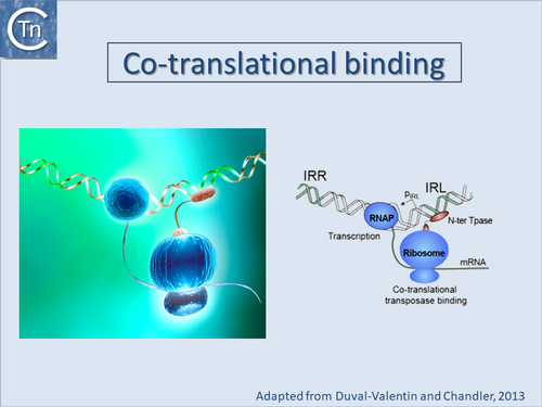
One biological explanation for cis preference is that the nascent N-ter domain might fold before completion of translation of the C-ter domain and the nascent protein could initiate binding directly to the closest IS end. Once bound, it would no longer be sensitive to masking by the C-ter domain. If binding fails to occur after translation of the N-ter DNA binding domain, continuing translation and folding of the C-ter domain would then mask the DNA binding domain resulting in an inactive protein. This implies that binding necessary for subsequent catalysis would occur only transitorily early in translation [[:File:1.36.1.png|(Fig.1.36.1)]]. | One biological explanation for cis preference is that the nascent N-ter domain might fold before completion of translation of the C-ter domain and the nascent protein could initiate binding directly to the closest IS end. Once bound, it would no longer be sensitive to masking by the C-ter domain. If binding fails to occur after translation of the N-ter DNA binding domain, continuing translation and folding of the C-ter domain would then mask the DNA binding domain resulting in an inactive protein. This implies that binding necessary for subsequent catalysis would occur only transitorily early in translation [[:File:1.36.1.png|(Fig.1.36.1)]]. | ||
| − | Direct evidence for co-translational binding was provided for IS''911'' using an in vitro transcription/translation system<ref><nowiki><pubmed>22195971</pubmed></nowiki></ref> where it was also demonstrated that reducing the efficiency of the -1 translational frameshifting required for IS''911'' transposase expression resulted in an increase in binding of a nascent transposase peptide. This is presumably because slowing the frameshifting process increases the time that the N-terminal part of the protein (which carries the sequence-specific DNA binding domain) is present on the ribosome enhancing its probability of binding to a neighboring IS end. It is interesting to note that in many IS, the DNA binding domain which recognizes the IR is located at the N-terminal end of the protein which is translated first. | + | Direct evidence for co-translational binding was provided for IS''911'' using an ''in vitro'' transcription/translation system<ref><nowiki><pubmed>22195971</pubmed></nowiki></ref> where it was also demonstrated that reducing the efficiency of the -1 translational frameshifting required for IS''911'' transposase expression resulted in an increase in binding of a nascent transposase peptide. This is presumably because slowing the frameshifting process increases the time that the N-terminal part of the protein (which carries the sequence-specific DNA binding domain) is present on the ribosome enhancing its probability of binding to a neighboring IS end. It is interesting to note that in many IS, the DNA binding domain which recognizes the IR is located at the N-terminal end of the protein which is translated first. |
One of the remaining questions concerns transposase multimerization. They must form multimers within the transpososome at some stage in the transposition pathway. Some transposases are monomeric in the absence of DNA (e.g. MuA and Tn''5''; <ref><nowiki><pubmed>1655409</pubmed></nowiki></ref><ref><nowiki><pubmed>9867814</pubmed></nowiki></ref><ref><nowiki><pubmed>18680433</pubmed></nowiki></ref>) while others are multimeric dimeric in solution (e.g. INHIV-1; <ref><nowiki><pubmed>12446721</pubmed></nowiki></ref>; <ref><nowiki><pubmed>17157316</pubmed></nowiki></ref>; <ref><nowiki><pubmed>19609359</pubmed></nowiki></ref>; <ref><nowiki><pubmed>19229293</pubmed></nowiki></ref>). | One of the remaining questions concerns transposase multimerization. They must form multimers within the transpososome at some stage in the transposition pathway. Some transposases are monomeric in the absence of DNA (e.g. MuA and Tn''5''; <ref><nowiki><pubmed>1655409</pubmed></nowiki></ref><ref><nowiki><pubmed>9867814</pubmed></nowiki></ref><ref><nowiki><pubmed>18680433</pubmed></nowiki></ref>) while others are multimeric dimeric in solution (e.g. INHIV-1; <ref><nowiki><pubmed>12446721</pubmed></nowiki></ref>; <ref><nowiki><pubmed>17157316</pubmed></nowiki></ref>; <ref><nowiki><pubmed>19609359</pubmed></nowiki></ref>; <ref><nowiki><pubmed>19229293</pubmed></nowiki></ref>). | ||
| Line 58: | Line 58: | ||
In view of the possibility that many transposases undergo co-translational binding, and the observation that several different purified full length transposases bind poorly to the ends of their cognate transposon (while the isolated N-terminal DNA binding domain alone binds robustly), it must be emphasized that purified transposases are probably largely inactive. This must be taken into account when assessing transposase properties. | In view of the possibility that many transposases undergo co-translational binding, and the observation that several different purified full length transposases bind poorly to the ends of their cognate transposon (while the isolated N-terminal DNA binding domain alone binds robustly), it must be emphasized that purified transposases are probably largely inactive. This must be taken into account when assessing transposase properties. | ||
| − | A recent study has provided support for the idea that transposases may also be able to multimerize cotranslationally. This study, has shown that bacterial luciferase subunits LuxA and LuxB may assemble cotranslationally | + | A recent study has provided support for the idea that transposases may also be able to multimerize cotranslationally. This study, has shown that bacterial luciferase subunits LuxA and LuxB may assemble cotranslationally i''n vivo.'' This process requires ribosome-associated trigger factor. This chaperone apparently delays subunit interactions until the LuxB dimer interface is available<ref><nowiki><pubmed>26405228</pubmed></nowiki></ref>. |
===Host factors=== | ===Host factors=== | ||
| Line 80: | Line 80: | ||
Accessory proteins: Acyl carrier protein (ACP), ribosomal protein L29, PepA and ArgR | Accessory proteins: Acyl carrier protein (ACP), ribosomal protein L29, PepA and ArgR | ||
| − | Acyl carrier protein (ACP) was independently shown to stimulate 3' end cleavage of Tn''3'' by its cognate Tpase<ref><nowiki><pubmed>9077464</pubmed></nowiki></ref> and, together with ribosomal protein L29, to greatly increase binding of TnsD (a protein involved in Tn''7'' target selection) to the chromosomal insertion site, attTn7<ref><nowiki><pubmed>16166383</pubmed></nowiki></ref>. Moreover ACP and L29 moderately stimulate Tn''7'' transposition in vitro while L29 alone has a significant stimulatory effect ''in vivo''<ref><nowiki><pubmed>16166383</pubmed></nowiki></ref>. The mode of action of these proteins may be similar to that of the accessory proteins PepA and ArgR which modify the architecture of the synaptic complex in certain XerC/XerD-mediated site-specific recombination reactions<ref><nowiki><pubmed>9348666</pubmed></nowiki></ref>. | + | Acyl carrier protein (ACP) was independently shown to stimulate 3' end cleavage of Tn''3'' by its cognate Tpase<ref><nowiki><pubmed>9077464</pubmed></nowiki></ref> and, together with ribosomal protein L29, to greatly increase binding of TnsD (a protein involved in Tn''7'' target selection) to the chromosomal insertion site, attTn7<ref><nowiki><pubmed>16166383</pubmed></nowiki></ref>. Moreover ACP and L29 moderately stimulate Tn''7'' transposition ''in vitro'' while L29 alone has a significant stimulatory effect ''in vivo''<ref><nowiki><pubmed>16166383</pubmed></nowiki></ref>. The mode of action of these proteins may be similar to that of the accessory proteins PepA and ArgR which modify the architecture of the synaptic complex in certain XerC/XerD-mediated site-specific recombination reactions<ref><nowiki><pubmed>9348666</pubmed></nowiki></ref>. |
<br /> | <br /> | ||
Revision as of 19:57, 5 May 2020
Contents
- 1 Transposase expression and activity
- 1.1 Impinging transcription
- 1.2 Sequestration of translation initiation signals
- 1.3 Programmed Translational Frameshifting
- 1.4 Programmed Transcriptional Frameshifting
- 1.5 Unusual amino acids Pyrrolysine and Selenocycteine
- 1.6 Translation termination
- 1.7 Transposase stability
- 1.8 Co-translational binding and multimerization
- 1.9 Host factors
- 2 Bibliography
Transposase expression and activity
While many of the classical mechanisms of controlling gene expression, such as the production of transcriptional repressors (IS1: [1][2]; IS2: [3] or translational inhibitors (anti-sense RNA in the case of IS10; see [4] are known to operate in Tpase expression, several other mechanisms have also been uncovered.
Impinging transcription
Many ISs have evolved mechanisms which attenuate their activation by impinging transcription following insertion into active host genes. This was originally observed in the case of IS1[5][6], for bacteriophage Mu[7] and IS50. To our knowledge other elements have not been examined.
This effect may be the result of disrupting complexes between Tpase and cognate DNA ends and could reflect either inhibition of transposase binding or disruption of extant transposase-end complexes.
In the case of bacteriophage Mu, transcription originating from within the element and impinging on the left end has also been shown to reduce activity[8]. It is possible that transcription disrupts the formation of intermediates including transposase and one or both Mu ends which lead to stable transposition complexes.
Sequestration of translation initiation signals
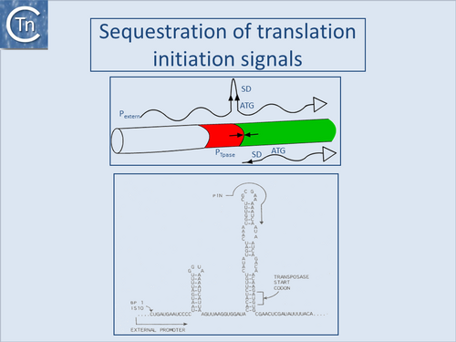
One such mechanism observed with IS10 and IS50, and potentially present in several other ISs, is the sequestering of translation initiation signals in an RNA secondary structure (Fig.1.32.1). These ISs carry inverted repeat sequences located close to the left end which include the ribosome binding site or translation initiation codon for the Tpase gene. Transcripts from the resident Tpase promoter include only the distal repeat unit which is unable to form the secondary structure, while transcripts from neighboring DNA include both repeats and would generate secondary structures in the mRNA which would sequester translation initiation signals[9][10]. This has been demonstrated experimentally for IS10 and IS50 but several additional insertion sequences carry such potential structures and might be expected to exhibit a similar mechanism (see [11]).
Programmed Translational Frameshifting
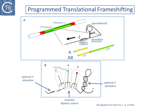
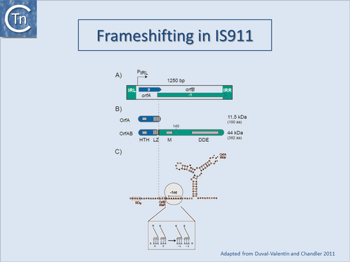
A second mechanism acts at the level of translation elongation and involves programmed translational frameshifting between two consecutive open reading frames (Fig.1.32.1). Typically a -1 frameshift is observed in which the translating ribosome slides one base upstream and resumes in the alternative phase. This generally occurs at the position of so-called slippery codons in a heptanucleotide sequence of the type X XXZ ZZN in phase 0 (where the bases paired with the anticodon are shown as triplets) which is read as XXX ZZZ N in the shifted -1 phase (Fig.1.32.1) (see e.g. [12][13][14], http://recode.genetics.utah.edu/). The sequence A AAA AAG is a common example of this type of heptanucleotide. Ribosomal shifting of this type is stimulated by structures in the mRNA which tend to impede the progression of the ribosome such as potential ribosome binding sites upstream or secondary structures (stem-loop structures and pseudoknots) downstream of the slippery codons. Translational control of transposition by frameshifting has been demonstrated both for IS1[15][16], and for members of the IS3 family (Fig.1.33.2) ([17]; see also [18]) but may also occur in several other IS elements (see for example IS5 and IS630 families). For IS1 and members of the IS3 family, the upstream frame appears to carry a DNA recognition domain whereas the downstream frame encodes the catalytic site. While the product of the upstream frame alone acts as a modulator of activity, presumably by binding to the IR sequences, frameshifting assembles both domains into a single protein, the Tpase, which directs the cleavages and strand transfer necessary for mobility of the element. The frameshifting frequency is thus critical in determining overall transposition activity. Although it has yet to be explored in detail, frameshifting could be influenced by host physiology thus coupling transposition activity to the state of the host cell.
Programmed Transcriptional Frameshifting
Another type of frameshifting may also occur in bacterial insertion sequences. This occurs at the transcriptional level and involves misreading and slippage of RNA polymerase on stretches of A residues. Although no real data is at present available, it may occur relatively frequently[19][20][21].
Unusual amino acids Pyrrolysine and Selenocycteine
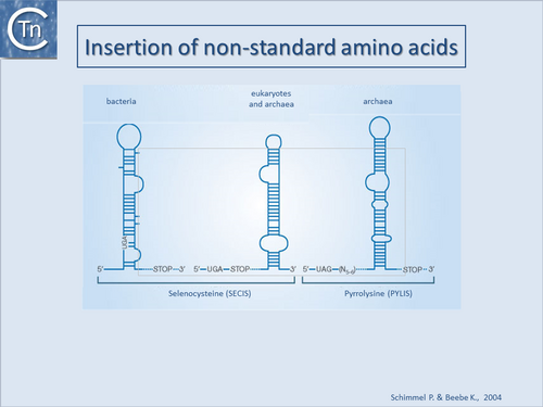
Another type of recoding which appears to occur in Methanosarcina, is the “suppression” of the stop codon, UAG, by insertion of Pyrrolysine. This was first noted in the Methylamine methyltransferases which are important in the production of methane by archaeal methanogens. [22] identified an in-frame amber codon (TAG) in the trimethylamine methyltransferase genes of both M. barkeri and M. thermophila. However, at least in the case of M. barkeri, abundant quantities of the full-length protein could be obtained and it appeared that the TAG codon was read as Lys. This later proved to be the unusual amino acid pyrrolysine. Several IS copies in these archaeal methanogens carry TAG codons which are presumably “suppressed” by decoding as pyrrolysine.
In this framework, other stop codons are known to be suppressed by decoding as selenocysteine [23]; [24][25][26]. To our knowledge a single IS from the IS3 family, ISDvu3 from Desulfovibrio vulgaris, includes a selenocysteine inserted at a stop codon in its orfB frame (M. Land personal communication). Undoubtedly additional examples will be identified in the future.
Inclusion of these amino acids involves the presence of specific types of secondary structures in the mRNA (Fig.1.33.3).
Translation termination
A third potential mechanism derives from the observation that the translation termination codon of Tpase genes of certain elements is located within their IR sequences. Although, to our knowledge, no extensive analysis of the significance of this arrangement has yet been undertaken, it seems possible that it may in some manner couple translation termination, transposase binding and transposition activity. The transposase gene of several elements does not possess a termination codon. These include IS240C, a member of the IS6 family (Y. Chen and J.Mahillon unpublished), two members of the IS5 family, IS427[27] and ISMk1[28] and various members of the IS630 family including IS870 and ISRf1[29]. Instead, some of these elements insert into a relatively specific target sequence in which the target DR produced on insertion itself generates the Tpase termination codon (see: IS630 family). The relevance of this as a control mechanism has yet to be explored.
Transposase stability
Transposase stability can also contribute to control of transposition activity. The Tpase of IS903 is sensitive to the E. coli Lon protease[30]. This sensitivity limits the activity of the Tpase both temporally and spatially and may provide an explanation for the observation that several Tpases function preferentially in cis. Indeed mutant IS903 Tpase derivatives have been isolated which exhibit an increased capacity to function in trans. These are more refractory to Lon degradation than the wildtype protein[31]. Some evidence that Lon may also be involved in regulating Tn5 (IS50) transposition has also been presented[32].
An observation which might also reflect Tpase instability is the temperature sensitive nature of IS1-mediated adjacent deletions in vivo[33], of Tn3 transposition[34] and of IS911 intramolecular recombination both in vivo and in vitro[35]. For IS911, incubation of the Tpase at 42°C results in an irreversible loss in activity. Further analyses showed that there was an increase in the proportion of transposase fragments some of which can presumably interact with full length transposase to inhibit its activity. This effect can be somewhat reduced by a series of mutation in the transposase gene whose function in stabilising the transposase is as yet unknown[36].
Co-translational binding and multimerization
Certain prokaryotic IS transposases show a strong preference for acting on the element from which they are expressed rather than on other copies of the same element in the cell. This phenomenon of “cis” preference presumably serves to prevent general activation of several identical IS copies by any “accidental” (stochastic) transposase expression from a single IS. Several different IS such as IS1[37], IS10[38], IS50[39], IS903[40] and IS911[41] (see [42][43] and references therein) exhibit this regulatory phenotype but “cis” preference may be the result of a combination of diverse mechanisms. Thus the Lon protease enhances “cis” preference of the IS903 transposase[44]. Transposition is enhanced in the absence of Lon and can be overcome by increased transposase expression[45]. For IS10, it is influenced by translation levels, Tpase mRNA half-life and translation efficiency[46][47].
Another mechanism, co-translational binding based on tight coupling between prokaryotic transcription and translation, was proposed to explain the inability to complement a Tpase mutant of IS903[48] and, more specifically, for Tn5[49].
Some full length IS transposases bind weakly to their cognate IR but the isolated DNA binding domain can bind more strongly. This has been observed for transposases of several elements including IS1[50] and IS30[51][52] and has also been observed for that of IS911. Early studies using band shift assays demonstrated that full length OrfAB binds the IRs only weakly and that OrfA binding was even lower or undetectable[53][54]. However, a truncated version of OrfAB, OrfAB [1-149], which is amputated for the C-terminal catalytic domain bound both ends avidly[55] (see also Transposase Stability). It is important to note this implies that, in many in vitro systems, the majority of transposase is thus likely to be inactive or only partially active since it would not bind stably to its substrate. The observations suggest that the C-terminal (C-ter) domain inhibits specific binding by the sequence-specific N-terminal DNA binding domain possibly by steric masking (Fig.1.36.1). This idea is consistent with the observation that IS10 transposase activity is increased by partial denaturation (for example by treatment with low alcohol concentrations; [56]). It is also consistent with the observation that the OrfAB protein of IS2 can bind the IS2 IRs when it carries a large GFP tag[57][58].
One biological explanation for cis preference is that the nascent N-ter domain might fold before completion of translation of the C-ter domain and the nascent protein could initiate binding directly to the closest IS end. Once bound, it would no longer be sensitive to masking by the C-ter domain. If binding fails to occur after translation of the N-ter DNA binding domain, continuing translation and folding of the C-ter domain would then mask the DNA binding domain resulting in an inactive protein. This implies that binding necessary for subsequent catalysis would occur only transitorily early in translation (Fig.1.36.1).
Direct evidence for co-translational binding was provided for IS911 using an in vitro transcription/translation system[59] where it was also demonstrated that reducing the efficiency of the -1 translational frameshifting required for IS911 transposase expression resulted in an increase in binding of a nascent transposase peptide. This is presumably because slowing the frameshifting process increases the time that the N-terminal part of the protein (which carries the sequence-specific DNA binding domain) is present on the ribosome enhancing its probability of binding to a neighboring IS end. It is interesting to note that in many IS, the DNA binding domain which recognizes the IR is located at the N-terminal end of the protein which is translated first.
One of the remaining questions concerns transposase multimerization. They must form multimers within the transpososome at some stage in the transposition pathway. Some transposases are monomeric in the absence of DNA (e.g. MuA and Tn5; [60][61][62]) while others are multimeric dimeric in solution (e.g. INHIV-1; [63]; [64]; [65]; [66]).
In view of the possibility that many transposases undergo co-translational binding, and the observation that several different purified full length transposases bind poorly to the ends of their cognate transposon (while the isolated N-terminal DNA binding domain alone binds robustly), it must be emphasized that purified transposases are probably largely inactive. This must be taken into account when assessing transposase properties.
A recent study has provided support for the idea that transposases may also be able to multimerize cotranslationally. This study, has shown that bacterial luciferase subunits LuxA and LuxB may assemble cotranslationally in vivo. This process requires ribosome-associated trigger factor. This chaperone apparently delays subunit interactions until the LuxB dimer interface is available[67].
Host factors
Transposition activity is frequently modulated by various host factors. These effects are generally specific for each element. A non-exhaustive list of such factors includes the DNA chaperones (or histone-like proteins), IHF, HU, HNS, and FIS, the replication initiator DnaA, the protein chaperone/proteases ClpX, P, and A, the SOS control protein LexA, and the Dam DNA methylase. In addition, proteins which govern DNA supercoiling in the cell can also influence transposition.
IHF, HU, HNS, and FIS
DNA chaperones may play roles in assuring the correct three dimensional architecture in the evolution of various nucleoprotein complexes necessary for productive transposition. They may also be involved in regulating Tpase expression. IHF, HU, HNS, and FIS have all been variously implicated in the case of bacteriophage Mu, in the control of Mu gene expression or directly in the transposition process (see [68] for review).
Several elements carry specific binding sites for IHF within, or close to, their terminal IRs. These can lie within (e.g. IS1: [69]; IS903: see [70] or close to (IS10: [71]) the Tpase promoter. IHF appears to influence the nature of IS10 transposition products by binding to a site 43 bp from one end [72], [73], [74].
IHF also stimulates Tpase binding to the ends of the Tn3 family member, Tn1000 or γδ [75]. Ironically, although IS1 was the first element in which IHF sites were identified (one within each IR), conditions have not yet been found in which IHF shows a clear effect on transposition or gene expression (D. Zerbib and M. Chandler, unpublished results).
The multiple roles of several of these proteins in both the IS10 and Tn5 (IS50) systems and the dynamics of their involvement has been determined in detail[76][77][78][79][80][81][82][83].
The histone-like nucleoid structuring protein H-NS, a global transcriptional regulator, has also been implicated in the regulation of bacterial transposition systems, including Tn10[84][85][86]. It appears to promote transposition by binding directly to the transposition complex (or transpososome).
DnaA
In the case of IS50, an element of the same family as IS10, both the protein Fis and the replication initiator protein DnaA have been reported to intervene in transposition (see [87]). Finally another "histone-like" protein, HNS, has been reported to stimulate transposition of IS1 in certain circumstances[88].
Although their mode of action is at present unknown, several other host proteins with otherwise entirely different functions have been implicated in transposition.
Accessory proteins: Acyl carrier protein (ACP), ribosomal protein L29, PepA and ArgR
Acyl carrier protein (ACP) was independently shown to stimulate 3' end cleavage of Tn3 by its cognate Tpase[89] and, together with ribosomal protein L29, to greatly increase binding of TnsD (a protein involved in Tn7 target selection) to the chromosomal insertion site, attTn7[90]. Moreover ACP and L29 moderately stimulate Tn7 transposition in vitro while L29 alone has a significant stimulatory effect in vivo[91]. The mode of action of these proteins may be similar to that of the accessory proteins PepA and ArgR which modify the architecture of the synaptic complex in certain XerC/XerD-mediated site-specific recombination reactions[92].
Bibliography
- ↑ <pubmed>2553980</pubmed>
- ↑ <pubmed>1848178</pubmed>
- ↑ <pubmed>8107136</pubmed>
- ↑ <pubmed>8556869</pubmed>
- ↑ <pubmed>6281761</pubmed>
- ↑ <pubmed>6313938</pubmed>
- ↑ <pubmed>3015876</pubmed>
- ↑ <pubmed>3015876</pubmed>
- ↑ <pubmed>2416461</pubmed>
- ↑ <pubmed>2438419</pubmed>
- ↑ <pubmed>7934941</pubmed>
- ↑ <pubmed>8384687</pubmed>
- ↑ <pubmed>8852897</pubmed>
- ↑ <pubmed>8811194</pubmed>
- ↑ <pubmed>2543983</pubmed>
- ↑ <pubmed>1848178</pubmed>
- ↑ <pubmed>1660923</pubmed>
- ↑ <pubmed>8384687</pubmed>
- ↑ <pubmed>PMC1088944</pubmed>
- ↑ <pubmed>16460832</pubmed>
- ↑ <pubmed>21673094</pubmed>
- ↑ <pubmed>10762254</pubmed>
- ↑ <pubmed>15788401</pubmed>
- ↑ <pubmed>23185002</pubmed>
- ↑ <pubmed>15372017</pubmed>
- ↑ <pubmed>12029131</pubmed>
- ↑ <pubmed>1963949</pubmed>
- ↑ <pubmed>8409920</pubmed>
- ↑ <pubmed>8387998</pubmed>
- ↑ <pubmed>2161528</pubmed>
- ↑ <pubmed>8898394</pubmed>
- ↑ <pubmed>2438419</pubmed>
- ↑ <pubmed>1101028</pubmed>
- ↑ <pubmed>378977</pubmed>
- ↑ <pubmed>9302015</pubmed>
- ↑ <pubmed>17078817</pubmed>
- ↑ <pubmed>6281761</pubmed>
- ↑ <pubmed>6299577</pubmed>
- ↑ <pubmed>6291787</pubmed>
- ↑ <pubmed>6271455</pubmed>
- ↑ <pubmed>22195971</pubmed>
- ↑ <pubmed>15207871</pubmed>
- ↑ <pubmed>2161528</pubmed>
- ↑ <pubmed>2161528</pubmed>
- ↑ <pubmed>8898394</pubmed>
- ↑ <pubmed>7692216</pubmed>
- ↑ <pubmed>8412678</pubmed>
- ↑ <pubmed>6271455</pubmed>
- ↑ <pubmed>6296834</pubmed>
- ↑ <pubmed>2826132</pubmed>
- ↑ <pubmed>15469518</pubmed>
- ↑ <pubmed>2154486</pubmed>
- ↑ <pubmed>10677279</pubmed>
- ↑ <pubmed>11352577</pubmed>
- ↑ <pubmed>9761671</pubmed>
- ↑ <pubmed>8132525</pubmed>
- ↑ <pubmed>22032517</pubmed>
- ↑ <pubmed>22277150</pubmed>
- ↑ <pubmed>22195971</pubmed>
- ↑ <pubmed>1655409</pubmed>
- ↑ <pubmed>9867814</pubmed>
- ↑ <pubmed>18680433</pubmed>
- ↑ <pubmed>12446721</pubmed>
- ↑ <pubmed>17157316</pubmed>
- ↑ <pubmed>19609359</pubmed>
- ↑ <pubmed>19229293</pubmed>
- ↑ <pubmed>26405228</pubmed>
- ↑ <pubmed>8805293</pubmed>
- ↑ <pubmed>2995832</pubmed>
- ↑ <pubmed>6261245</pubmed>
- ↑ <pubmed>8556869</pubmed>
- ↑ <pubmed>7744253</pubmed>
- ↑ <pubmed>9630232</pubmed>
- ↑ <pubmed>7556079</pubmed>
- ↑ <pubmed>2844529</pubmed>
- ↑ <pubmed>9630232</pubmed>
- ↑ <pubmed>11447129</pubmed>
- ↑ <pubmed>14530435</pubmed>
- ↑ <pubmed>14992723</pubmed>
- ↑ <pubmed>19696075</pubmed>
- ↑ <pubmed>19042975</pubmed>
- ↑ <pubmed>16166383</pubmed>
- ↑ <pubmed>17501923</pubmed>
- ↑ <pubmed>19696075</pubmed>
- ↑ <pubmed>16166383</pubmed>
- ↑ <pubmed>17501923</pubmed>
- ↑ <pubmed>7504907</pubmed>
- ↑ <pubmed>10583504</pubmed>
- ↑ <pubmed>9077464</pubmed>
- ↑ <pubmed>16166383</pubmed>
- ↑ <pubmed>16166383</pubmed>
- ↑ <pubmed>9348666</pubmed>
